InfraLife Exchange dialogue:
Imaging
Exchange dialogue – Imaging
During the spring of 2023, InfraLife arranges exchange dialogues on Imaging. There are several state-of-the-art opportunities within imaging at the three infrastructures. Several beamlines at MAX IV offer X-ray techniques relevant for imaging, such as tomography and X-ray fluorescence. Once ESS is up and running, there will also be several beamlines offering imaging using neutrons. At SciLifeLab there are a number of units offering imaging techniques, e.g., Advanced Light Microscopy, Advanced FISH Technologies and Cryo EM.
Below is a preliminary schedule for the first three sessions.
If you are interested in participating in these sessions, please josefin.lundgren.gawell@scilifelab.se
Session #4: SciLifeLab AIDA Data Hub
Date: May 2, Time: 11.00-12.00, Location: Zoom
AIDA Data Hub – clinical imaging for AI research in SciLifeLab
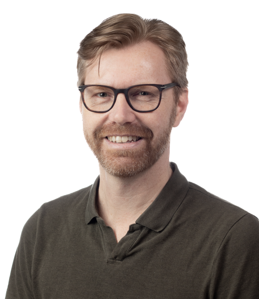
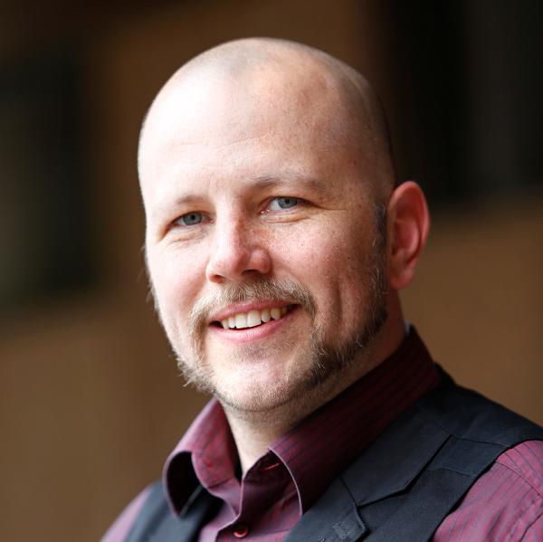
Claes Lundström (Platform Scientific Director, NBIS) and Joel Hedlund (Data director CMIV/AIDA, AIDA Data Hub Lead) will be presenting.
AI research has vast potential to make great advances for healthcare, not the least in diagnostic imaging. However, effective access to realistic clinical data is difficult for several reasons, a primary one being that it is sensitive personal data. The AIDA Data Hub provides services to enable and accelerate healthcare-native AI research.
“The AIDA Data Hub offers Data Sharing, Policy Support, and Services for the Swedish medical imaging diagnostics AI research community. The purpose is to gather, annotate, and share large volumes of data necessary in order to enable research, innovation and clinical adoption of world-class AI technology. We also operate a GPU compute system for AI training on sensitive personal data for Swedish researchers”, says xx.
More information
Session #3: Imaging with neutrons, presented by representatives from European Spallation Source (ESS)
Date: April 18, Time: 11.00-12.00, Location: Zoom
Neutron imaging @ESS, principles and applications
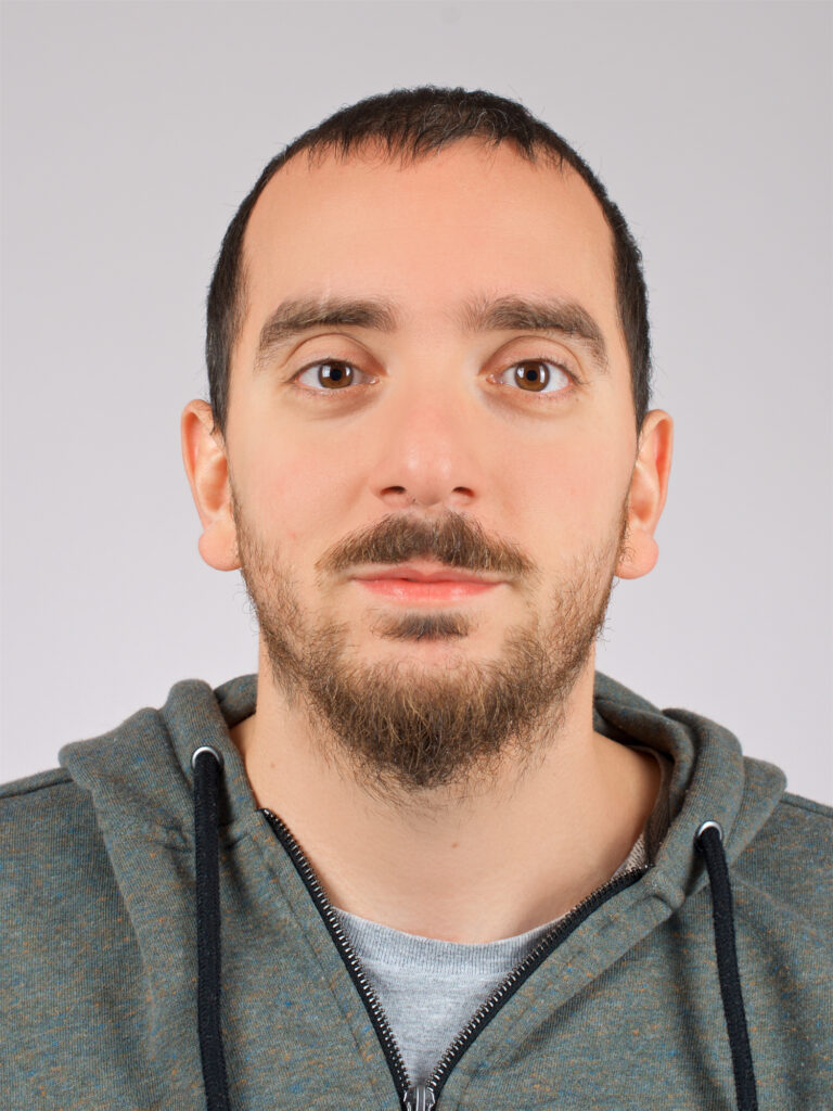
Manuel Morgano (instrument scientist at ESS Odin) will give an introduction to lorem ipsum sit dolor amet.
“Neutron imaging so far has been a “niche” technique, but it is now becoming more and more mainstream due to the widespread recognition of its capabilities. With ODIN @ESS, our aim is to make it easily available and flexible to a wide range of users, both in academia and industry”, Manuel says.
More information
Session #2: Imaging at MAX IV – Examples from the ForMAX, SoftiMAX and NanoMAX beamlines
Date: April 4, Time: 11.00-12.00, Location: Zoom
Going subcellular at SoftiMAX & NanoMAX: Life Science examples at X-ray Nanoprobes”
Karina Thånell (Group Manager of Imaging: SoftiMAX, NanoMAX, MAXPEEM, STM) will give an introduction to X-ray nanoprobes at SoftiMAX and NanoMAX.
“I’m curious to see the potential for ‘multimodal approaches’ at other facilities, as well as data analysis tools for the users”, Karina says.

Full-field tomographic imaging at ForMAX
Samuel McDonald (Scientist at ForMAX beamline) will walk us through the process of full-field tomographic imaging.
“I am interested to hear about image processing pipelines and data analysis tools and procedures that other infrastructures provide”, says Samuel.
Mapping nanoscale structures with scanning SWAXS imaging
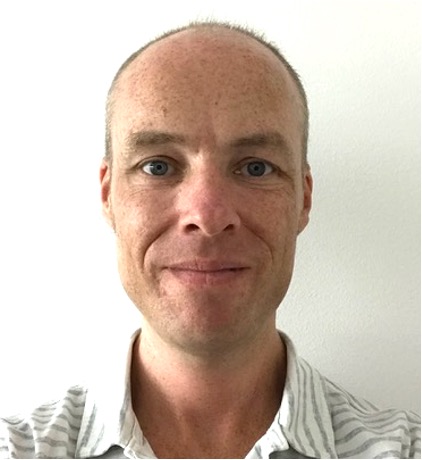
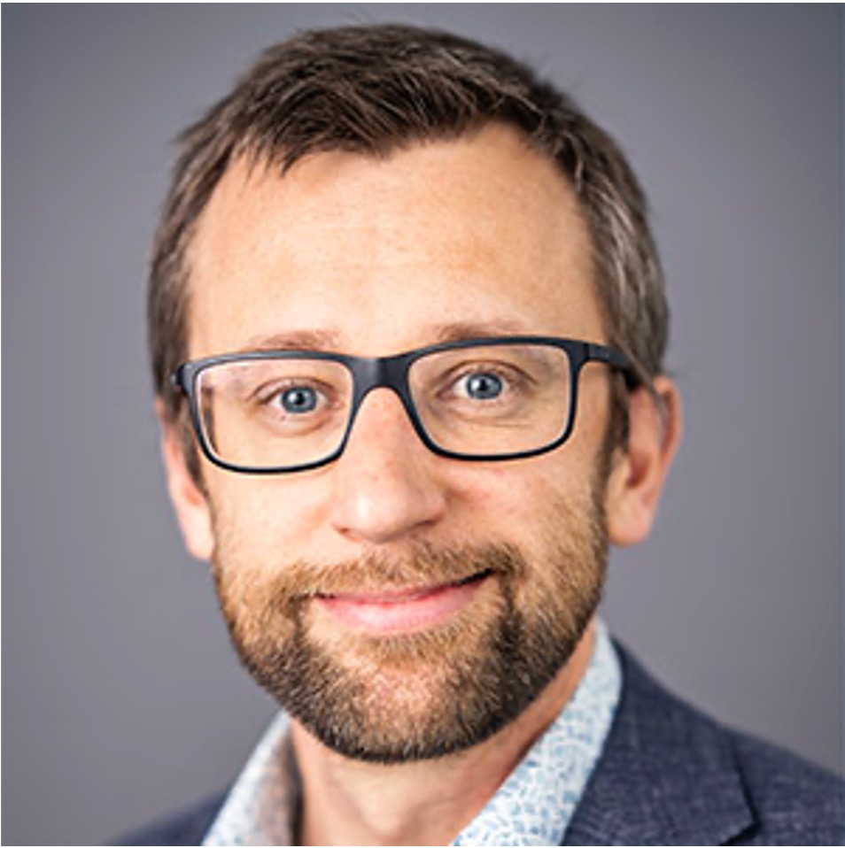
Kim Nygård, Beamline Manager at ForMAX beamline, will walk us through the process of mapping nanoscale structures using SWAXS imaging. Perspiciatis unde omnis iste natus error sit voluptatem alop?
“I’d like to find out what complementary techniques the other infrastructures provide”, says Kim.
More information
Session #1: Imaging and imaging data analysis at SciLifeLab
Date: March 14, Time: 11.00-12.00, Location: Zoom
Unique microscopy imaging opportunities distributed throughout Sweden – at SciLifeLab Cell and Molecular Imaging Platform (CMI) and the National Microscopy Infrastructure nodes (NMI, a VR research infrastructure)
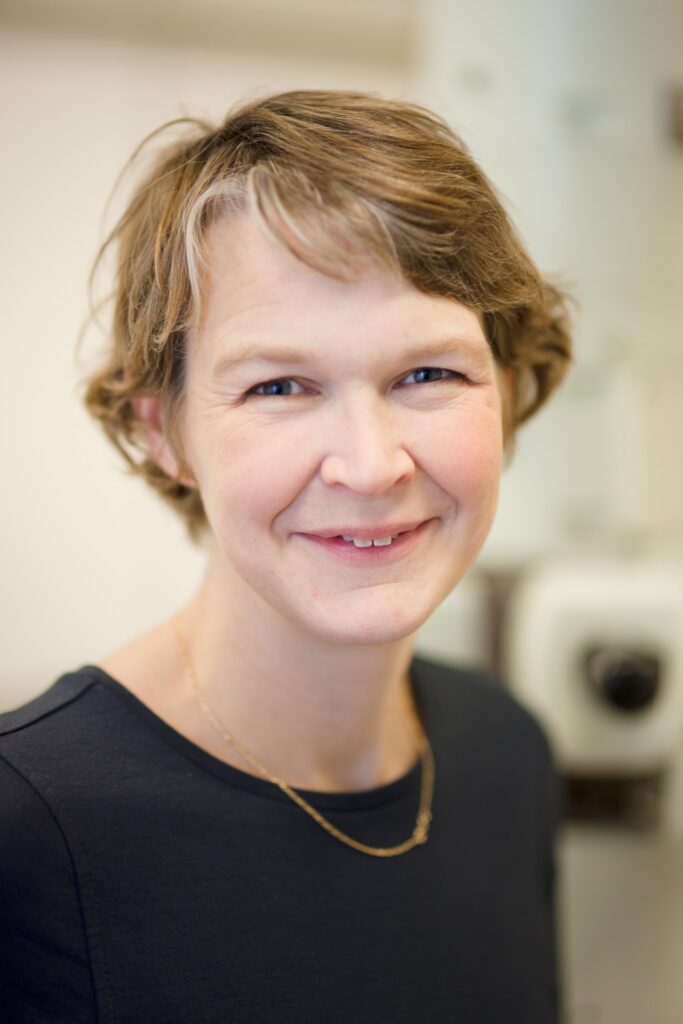
Linda Sandblad (Director of Umeå Centre for Electron Microscopy (UCEM) and Scientific Director at CMI) will take us through sed ut perspiciatis unde omnis iste natus error sit voluptatem accusantium doloremque laudantium, totam rem aperiam.
“Sample preparation is an important step that influence image quality and data interpretations when using electron microscopy. I am curious about what type of preparation is best for other imaging methods and how it influences analysis for these techniques”, says Linda.
BioImage Informatics Facility (BIIF) – image analysis support in Sweden
Anna Klemm (Head of BioImage Informatics Facility, SciLifeLab; Uppsala University), will give an introduction to sed ut perspiciatis unde omnis iste natus error sit voluptatem accusantium doloremque laudantium.
“I look forward to learning more about what kind of imaging data other infrastructures produce, where images are stored and how they are analysed. Also, if there are standard open-source software tools or pipelines available for the image data, what are those?”, says Anna.
More information
Linda Sandblad’s presentation
Anna Klemm’s presentation
Other useful links highlighted at the session
Sign up for the Digital image analysis for scientific applications course (2023-04-11 to 2023-05-09).
5th NEUBIAS Conference in Porto, Portugal 8 – 12 May 2023
COMULIS (Correlated Multimodal Imaging in Life Sciences)
A Hitchhiker’s guide through the bio-image analysis software universe
PRISMAS – PhD Research and Innovation in Synchrotron Methods and Applications in Sweden
Do you want to take part in upcoming meetings or have a suggestion for future sessions? Get in touch!